
Urinary Sediment Stain (Modified S&M)
Intended use:
Urinary analysis is one of the three routines in clinical examination and widely used in clinic. Urinary sediment test, as the most common examination for urinary analysis, is mainly used to identify pathological components in urine including cells, casts, crystal, bacteria and parasites. It is of great value for diagnosis, orientation, identification and prognosis estimation of urinary system disease.
Principle:
SM Urinary sediment stain is the most widely-used method. Directly add stain into urinary sediment, and the components in urinary sediment can be easily identified based on their distinct shape and structure. Compared with the traditional SM stain, this modified BASO Urinary sediment stain offers the benefits of shorter staining time and longer storage stability at room temperature.
Specifications:
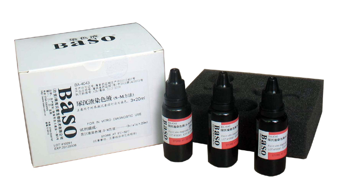
| Contents | 3vialsx20ml/kit | Components |
| Urinary sediment stain | 20ml | Gentian Violet, Ammonium Oxalate, Ethanol, Safranin O |
Expected Results:
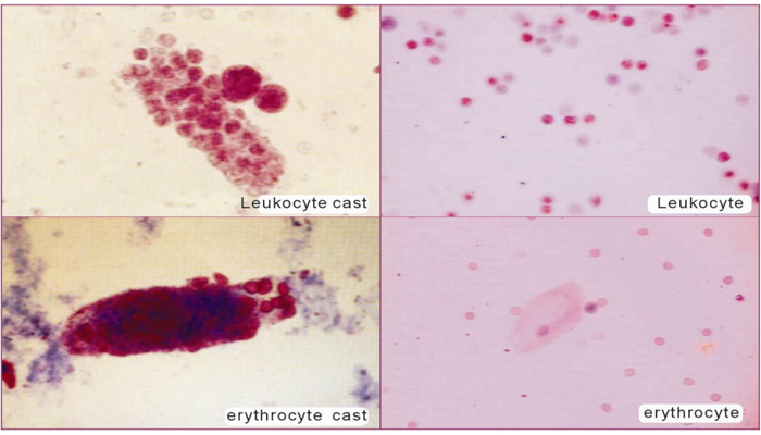
1. Neutrophilic leukocytes are stained purple; nuclei are stained red-purple.
2. Vaginal squamous epithelial cells are stained pale purple; nuclei are stained dark purple.
3. Bladder epithelial cells are colorless or pale blue.
4. Hyaline casts are stained pink or light purple.
5. Granules are stained red and purple; Granular castsare stained blue.
6. Cytoplasm of lipocyte is lightly stained, with visible alveolate structure.
7. Living and active bacteria are stained pink, dead bacteria are stained dark purple.
8. Cells of yeasts are stained dark purple or unstained.
9. Trichomonas are stained colorless or pale blue.
10. Erythrocytes are stained light red-purple.